Media Summary: MIT 7.016 Introductory Biology, Fall 2018 Instructor: Adam Martin View the complete course: Dr. Mathieu Frechin, Head of Quantitative Biology at Nanolive will introduce you to our holotomographic Real-Time IntraVital Microscopy (IVM): In Vivo

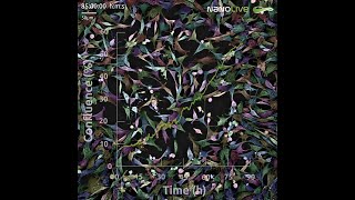
Webinar 4d Live Cell Imaging - Detailed Analysis & Overview

MIT 7.016 Introductory Biology, Fall 2018 Instructor: Adam Martin View the complete course: Dr. Mathieu Frechin, Head of Quantitative Biology at Nanolive will introduce you to our holotomographic Real-Time IntraVital Microscopy (IVM): In Vivo Are you looking for something to do over the festive period? Why not watch our latest Light Microscopy - Fundamental Principles - Presented By: Jun Park, PhD Speaker Biography: Jun Park, Ph. D. is Senior Scientist and Applications Lead at MilliporeSigma.

Photo Gallery

Webinar 4D Live Cell Imaging
Five steps for publication-quality fixed-cell imaging the first time Complete Webinar
Live Cell Imaging Webinar | How to monitor and quantify cell morphology
[Webinar] Maintaining Cell Health with Live-cell Imaging
[Webinar] Real-time monitoring and analysis of 3D cell models with live-cell imaging solutions
Live Cell Imaging Webinar | How to track and quantify cell movement
29. Cell Imaging Techniques
Webinar | Label-free live cell imaging meets Immuno-Oncology
(EN) Webinar: Real-time Intravital Microscopy for Biomedical Research
Best practices: 5 steps to live-cell imaging
Live Cell Imaging Webinar | Introduction to label-free live cell imaging
Live Cell Imaging Webinar | Reveal your cells with 1 experiment and 8 applications
Sponsored
Sponsored
View Detailed Profile
Webinar 4D Live Cell Imaging

Webinar 4D Live Cell Imaging

Webinar

Five steps for publication-quality fixed-cell imaging the first time Complete Webinar

Five steps for publication-quality fixed-cell imaging the first time Complete Webinar

https://www.thermofisher.com/us/en/home/life-science/

Sponsored
Live Cell Imaging Webinar | How to monitor and quantify cell morphology

Live Cell Imaging Webinar | How to monitor and quantify cell morphology

In this

[Webinar] Maintaining Cell Health with Live-cell Imaging

[Webinar] Maintaining Cell Health with Live-cell Imaging

Maintaining Cell Health with

[Webinar] Real-time monitoring and analysis of 3D cell models with live-cell imaging solutions

[Webinar] Real-time monitoring and analysis of 3D cell models with live-cell imaging solutions

Discover the Future of 3D

Sponsored
Live Cell Imaging Webinar | How to track and quantify cell movement

Live Cell Imaging Webinar | How to track and quantify cell movement

In this

29. Cell Imaging Techniques

29. Cell Imaging Techniques

MIT 7.016 Introductory Biology, Fall 2018 Instructor: Adam Martin View the complete course: https://ocw.mit.edu/7-016F18 ...

Webinar | Label-free live cell imaging meets Immuno-Oncology

Webinar | Label-free live cell imaging meets Immuno-Oncology

Dr. Mathieu Frechin, Head of Quantitative Biology at Nanolive will introduce you to our holotomographic

(EN) Webinar: Real-time Intravital Microscopy for Biomedical Research

(EN) Webinar: Real-time Intravital Microscopy for Biomedical Research

Real-Time IntraVital Microscopy (IVM): In Vivo

Best practices: 5 steps to live-cell imaging

Best practices: 5 steps to live-cell imaging

Webinar

Live Cell Imaging Webinar | Introduction to label-free live cell imaging

Live Cell Imaging Webinar | Introduction to label-free live cell imaging

In this

Live Cell Imaging Webinar | Reveal your cells with 1 experiment and 8 applications

Live Cell Imaging Webinar | Reveal your cells with 1 experiment and 8 applications

During this

Video4 hypoxia dye live cell imaging

Video4 hypoxia dye live cell imaging

Video4 hypoxia dye live cell imaging

Label-free live cell imaging phenotypic variation in single cells. Webinar now available on demand!

Label-free live cell imaging phenotypic variation in single cells. Webinar now available on demand!

Are you looking for something to do over the festive period? Why not watch our latest

Technical Tea Break - Live Cell Imaging, Nadia Halidi

Technical Tea Break - Live Cell Imaging, Nadia Halidi

Light Microscopy - Fundamental Principles -

Incredible 4D live imaging of T-cell nuclear dynamics

Incredible 4D live imaging of T-cell nuclear dynamics

4D live imaging

Best practices: 5 steps to live-cell imaging

Best practices: 5 steps to live-cell imaging

https://www.thermofisher.com/us/en/home/life-science/

Dynamic live cell imaging of adherent and immune cells using CellASIC® ONIX2 microfluidic platform

Dynamic live cell imaging of adherent and immune cells using CellASIC® ONIX2 microfluidic platform

Presented By: Jun Park, PhD Speaker Biography: Jun Park, Ph. D. is Senior Scientist and Applications Lead at MilliporeSigma.