Media Summary: In this video - obtained with Nanolive's 3D Dr. Mathieu Frechin, Head of Quantitative Biology at Nanolive will introduce you to our holotomographic DMU students are among the first undergrad students in the world to use
Label Free Live Cell Imaging - Detailed Analysis & Overview
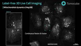
In this video - obtained with Nanolive's 3D Dr. Mathieu Frechin, Head of Quantitative Biology at Nanolive will introduce you to our holotomographic DMU students are among the first undergrad students in the world to use The video features primary human keratinocytes isolated from the epidermis of human skin. The function of keratinocytes is to ... Mitochondria are highly dynamic organelles that maintain their morphology via continuous fission and fusion, also known as ... You asked – we delivered! A complete solution for automated, quantitative,