Media Summary: DMU students are among the first undergrad students in the world to use Mitochondria are highly dynamic organelles that maintain their morphology via continuous fission and fusion, also known as ... On the left panel you can observe a time-lapse of pre-adipocytes imaged with the
3d Label Free Live Cell - Detailed Analysis & Overview
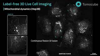
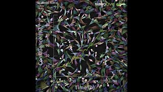
DMU students are among the first undergrad students in the world to use Mitochondria are highly dynamic organelles that maintain their morphology via continuous fission and fusion, also known as ... On the left panel you can observe a time-lapse of pre-adipocytes imaged with the Dr. Mathieu Frechin, Head of Quantitative Biology at Nanolive will introduce you to our holotomographic microscopy and its ... In this movie (killing assay) you can see T- A time-lapse video of a Chinese Hamster Ovary (CHO)
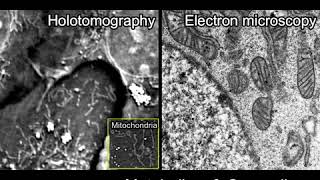
Conventional Imaging Tools vs Check out this insightful animation explaining the constant struggles with ... The video features primary human keratinocytes isolated from the epidermis of human skin. The function of keratinocytes is to ... Are you looking for something to do over the festive period? Why not watch our latest webinar about phenotypic variation in single ... This video shows a loss of mitochondrial potential (fluorescence intensity is progressively reducing) over the time of the movie.